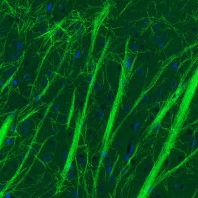

Although its exact role remains unclear, it is thought to act as a cellular adhesive molecule, to be involved as a regulator of oligodendrocyte microtubule stability and to mediate complement cascade.

Myelin oligodendrocyte glycoprotein (MOG) is a glycoprotein located on the myelin surface and found exclusively in the central nervous system (CNS). Randomised control trials are limited, but observational open-label experience suggests a role for high-dose steroids and plasma exchange in the treatment of acute attacks, and for immunosuppressive therapies, such as steroids, oral immunosuppressants and rituximab as maintenance treatment. Cerebrospinal fluid oligoclonal bands are uncommon. MRI characteristics can help in differentiating MOG-AD from other neuroinflammatory disorders, including multiple sclerosis and neuromyelitis optica. To avoid misdiagnosis, MOG antibody testing should be undertaken in selected cases presenting clinical and paraclinical features that are felt to be in keeping with MOG-AD, using a validated cell-based assay. Recent advances in MOG antibody testing offer improved sensitivity and specificity. Residual disability develops in 50–80% of patients, with transverse myelitis at onset being the most significant predictor of long-term outcome. Disease course can be either monophasic or relapsing, with subsequent relapses most commonly involving the optic nerve. Although no age group is exempt, median age of onset is within the fourth decade of life, with optic neuritis being the most frequent presenting phenotype. Study limitations include the variability in treatments used, which prevented the researchers from determining which immune lowering treatment was best.Myelin oligodendrocyte glycoprotein (MOG) antibody disease (MOG-AD) is now recognised as a nosological entity with specific clinical and paraclinical features to aid early diagnosis. In 2017, Mayo Clinic developed the MOG antibody test, the first of its kind in the United States. Dubey says, because its presence helps predict patients who are more likely to relapse. Testing for the MOG antibody also helps from a prognostic standpoint, Dr. This should be dark grey throughout, and you can see some white area that forms a 'H-sign' appearance, which is what we frequently see with the MOG antibody." - Dr. Image A2: "The oval grey circle is the spinal cord in transverse plane (vertical plane dividing body top from bottom through waist) indicated by the arrow. “Patients who have the MOG antibody get inflammation that damages this insulation, with loss of signaling in the spinal cord resulting in paralysis,” says Divyanshu Dubey, M.B.B.S., a Mayo Clinic neurologist and first author of the article. The nerves of the spinal cord are covered with an insulating layer called the myelin sheath, similar to insulation of an electrical wire. “It is important for physicians to search for this MOG antibody in individuals who present with paralysis from spinal cord inflammation because the patients with MOG antibody respond well to treatment that lowers the immune system response,” says Eoin Flanagan, M.B., B.Ch., a Mayo Clinic neurologist and senior author of the article. Patients with MOG antibodies may respond to steroids and additional treatments, such as plasma exchange to remove these antibodies from the blood, but acute flaccid myelitis does not respond well to such treatments. Of 54 patients in the study, 19 percent with spinal cord inflammation from the MOG antibody were initially misdiagnosed with acute flaccid myelitis. MOG is a helpful biomarker of transverse myelitis, the researchers report. The mimicry finding was an unexpected discovery, as the study focused on patients with transverse myelitis, a disease of spinal cord inflammation that also can lead to paralysis. It should be dark grey throughout ,and this shows a bright line (arrowhead) surrounded by more hazy white signal (arrows) that we often see with MOG antibody cases." - Dr. The cervical spinal cord is the grey structure the arrows are pointing to, and it should be dark grey like it is at the top of the image. Image A1: "A view of the neck in sagittal plane (as if a line was drawn down the a person's middle top to bottom between eyes). The researchers show that detecting the MOG antibody has important treatment and prognostic implications. Mayo Clinic researchers report that spinal cord inflammation associated with an antibody to myelin oligodendrocyte glycoprotein (MOG) can mimic acute flaccid myelitis, a rare but serious disease linked to certain viruses that particularly affects children and can result in paralysis.


 0 kommentar(er)
